See More. Detect More. Do More.​

Pristine 5.0​

A next-generation widefield retinal camera that delivers compact, high-resolution, true-color retinal imaging for precise photo-documentation of peripheral lesions and retinal diseases.​

Serves a

~USD

370.6

Million

Growing at a CAGR of 6.4 %​

Fundus Camera Market.​

Pristine 5.0 serves the diagnostic retinal imaging market, supporting routine clinical diagnosis in high-throughput diagnostic environments as well as long-term monitoring of chronic retinal disease. ​Rising chronic disease burden has resulted in the need for a workflow-efficient,  affordable and ​automated widefield imaging system​

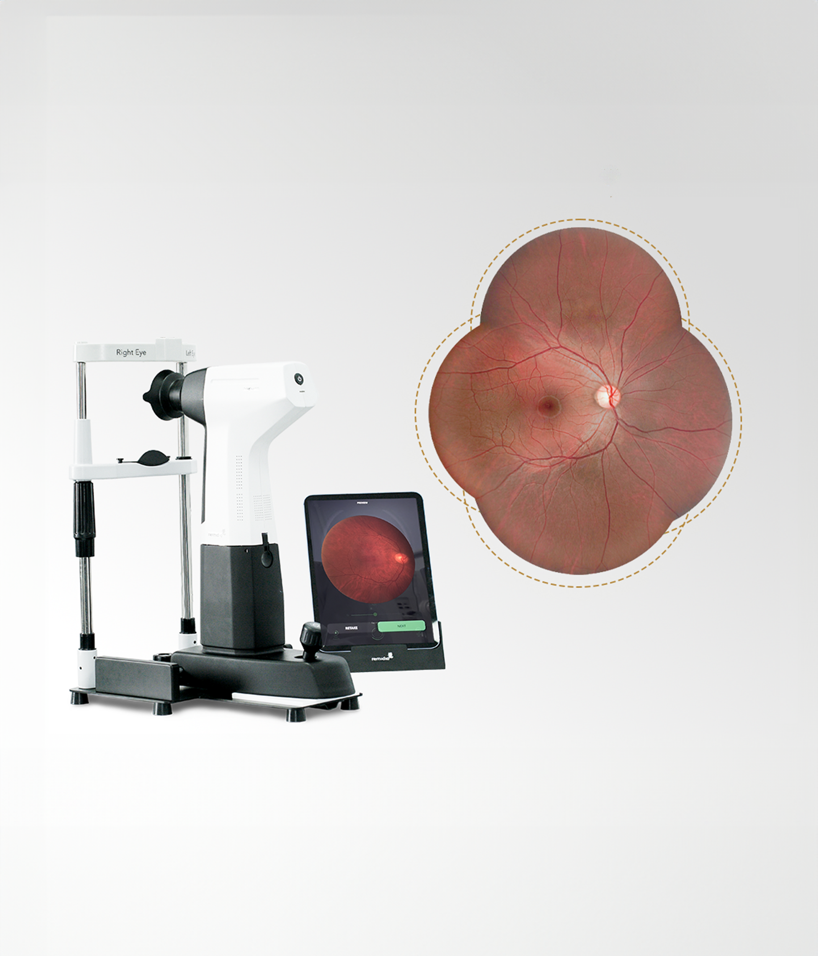
High-Resolution Widefield Retinal Imaging for Routine Care​

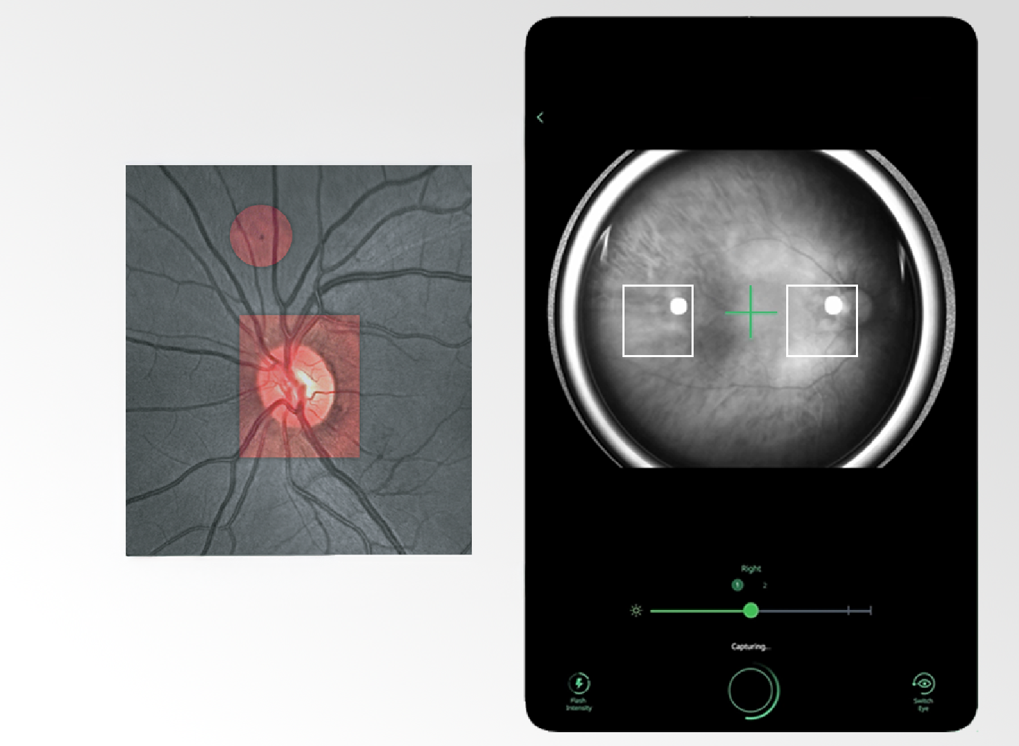
Conventional fundus cameras miss peripheral pathologies, while ultra-wide field systems are bulky, expensive, complex, lack true-color imaging, and are impractical for routine use.​

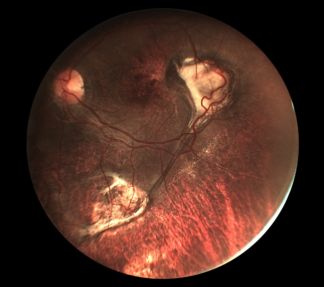
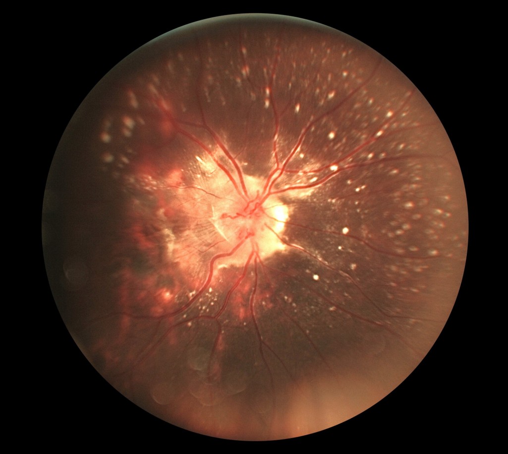
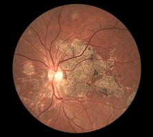
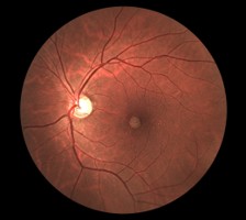
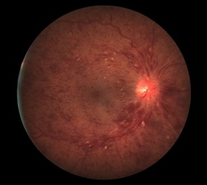
Clinical evidence shows that an ETDRS 7-field view (~75°) is sufficient to detect most sight-threatening retinal disease, enabling earlier identification of peripheral ischemia, timely intervention, and improved long-term outcomes.​

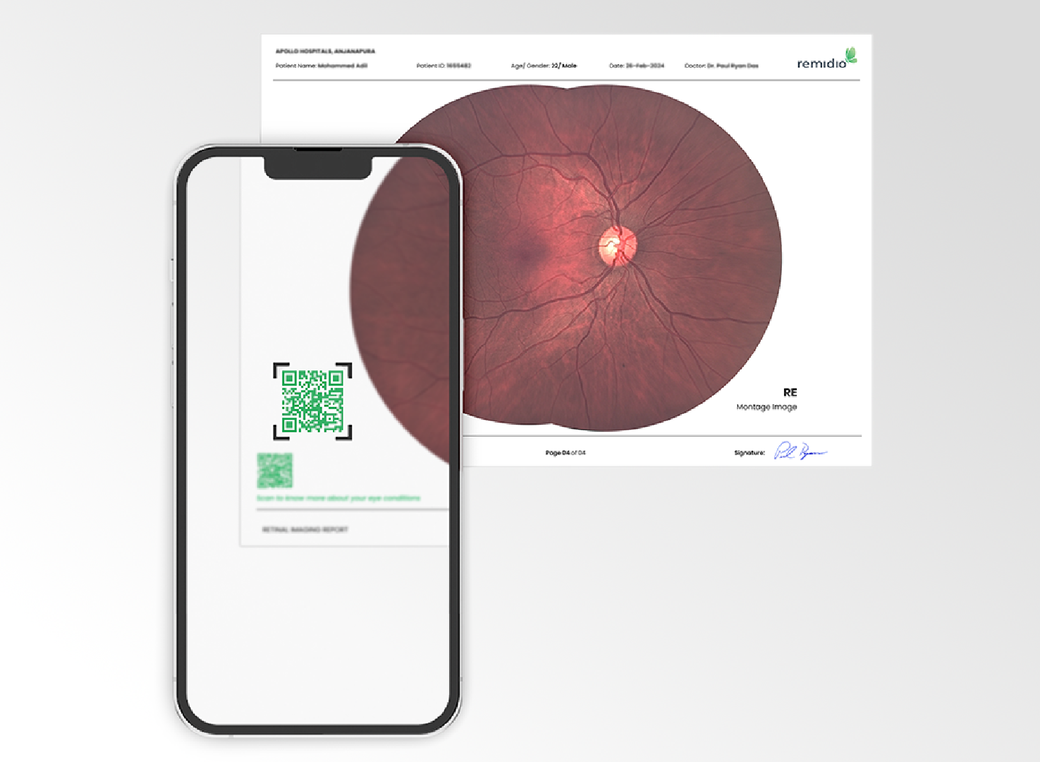
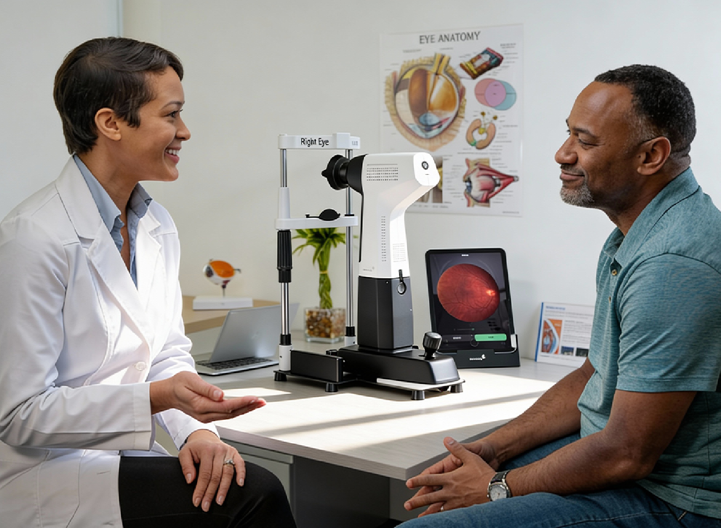
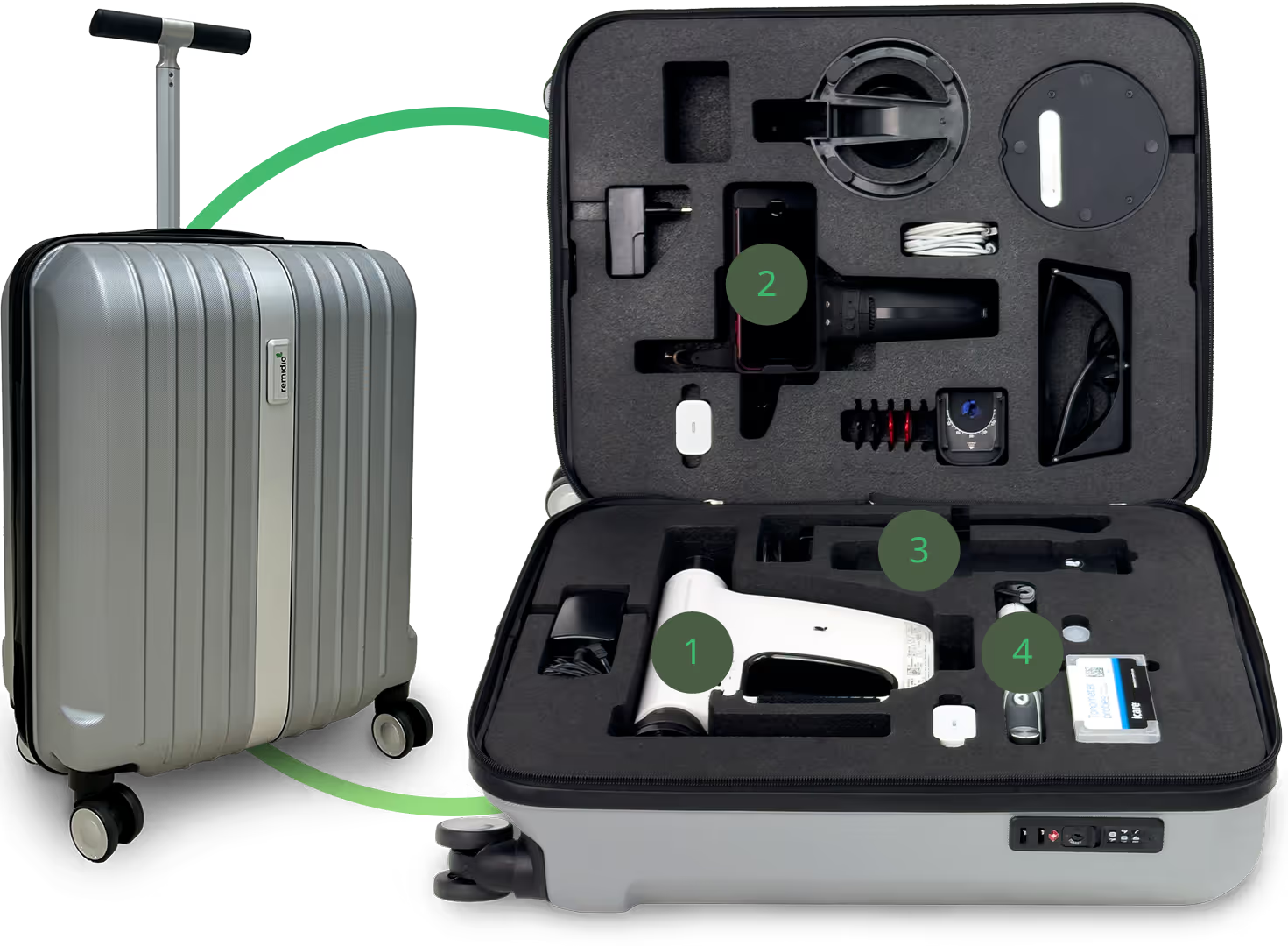
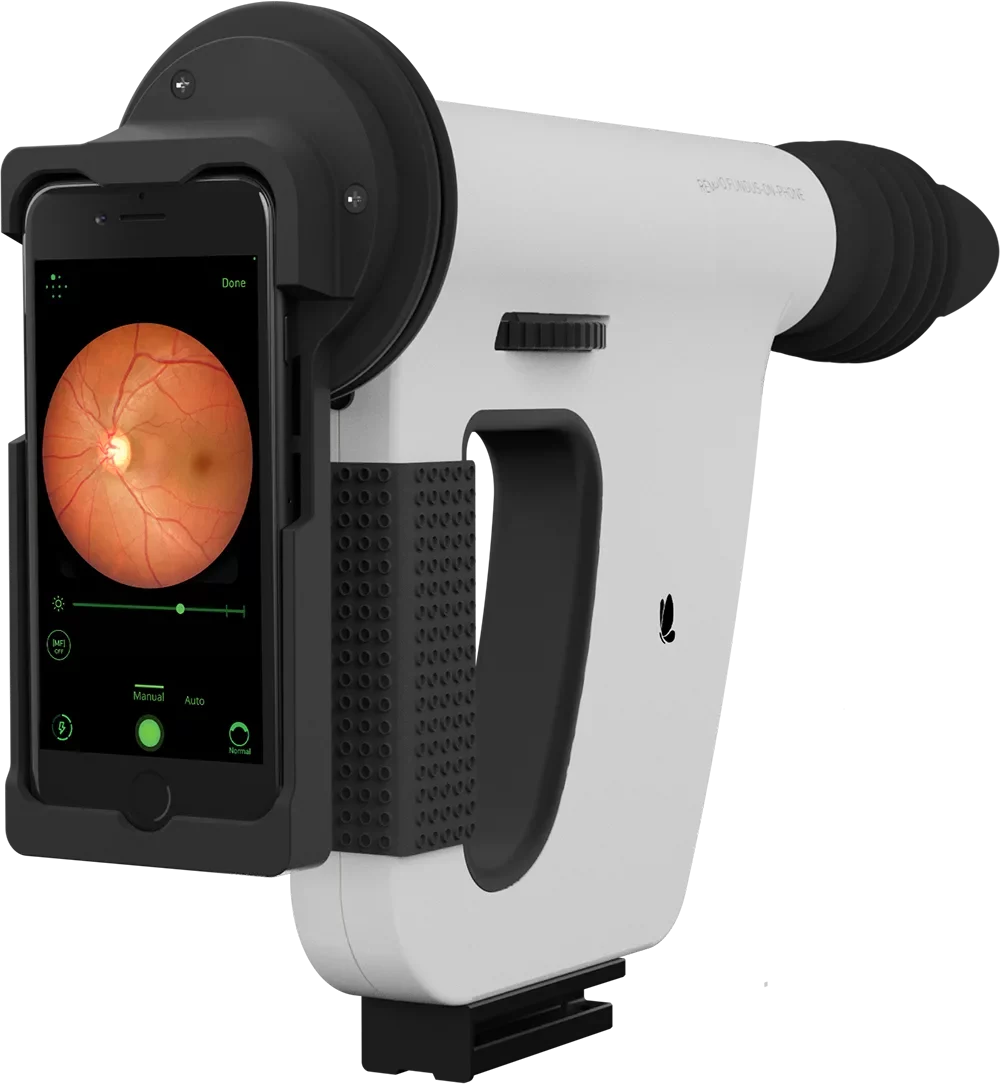
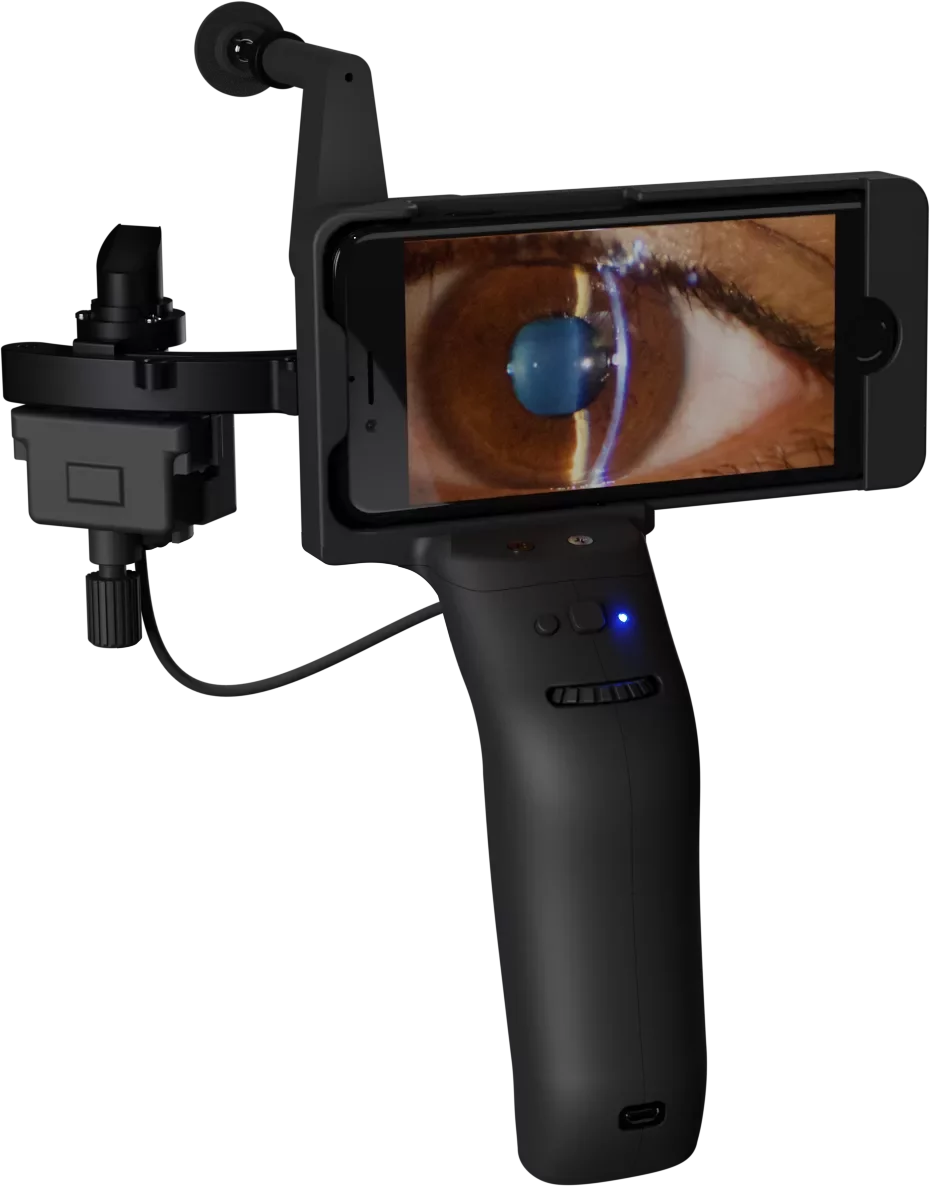
Pristine 5.0 represents a breakthrough in affordable, high-resolution, true-color widefield imaging.​​

Patent

Reflex Free Novel Illumination Design​

Annular Co-axial Illumination Technology
Eliminates Corneal Reflex Artefacts by using
black spot / absorbers and separating illumination and imaging paths​.

Product Features

A space-saving widefield imaging system with a movable iPad interface that supports easy setup and enables clear, visual conversations with patients on acquired images​.
Delivers high-resolution images with auto-focus and auto-capture, making retinal imaging fast, accurate, and easy for everyday clinical use​.
Engage, empower & educate patients via patented QR- coded reports that unlock interactive awareness videos and critical information on retinal health.​
Low-cost maintenance, and seamless workflow integration with DICOM and DICOM Web compatibility, delivering a smooth experience for both clinics and patients.​

Smart Solutions Tailored
for Every Setting

Medical Retina Practices for photo
documentation pre and post treatment​

Vitreo-retinal clinics for high quality
photo documentation and follow up​

General Ophthalmology practices as
an annual screening device for peripheral
pathologies​

Cornea refractive practices for screening
for myopic degeneration as well as for
lattices and holes​

Clinical Validation & Device Performance​

Images captured on Pristine 5.0

Fundus images captured on Pristine 5.0 by our customers and in-house optometrists.

Specifications

Type of imaging​​

Mydriatic

Resolution​​

20.2 Megapixel​​

Normal Mode Fixation​​

2 Internal fixation (Disc, Macula ) ​​

Expert Mode Fixation​​

4 Internal fixation (Disc, Macula, Superior, Inferior) ​​

Image Mode​​

Color, Red Free,
Anterior Diffused,
Posterior Reflex free imaging​​

Minimum Pupil Size ​​

5mm or Greater​​

Capture Mode Posterior​​

Auto focus and Auto Capture​​

Field of View In Single Short ​​

Typical 58o

Expert Mode Fixation​​

4 Internal fixation (Disc, Macula, Superior, Inferior) ​​

Montage​​

2 Image (87o x 58°) , 4 Image (87o x 116o)​​

Working distance for retinal imaging ​​

18 mm​​

Refractive error range​​

+20 / -20 Dioptre ​​

Screen size​​

10.9 inch​​
Download brochure

What our Customers say

Product Video

Regulatory Approvals

CDSCO
Approved